Omaha:
(402) 496-9733
Bellevue:
(402) 291-0528


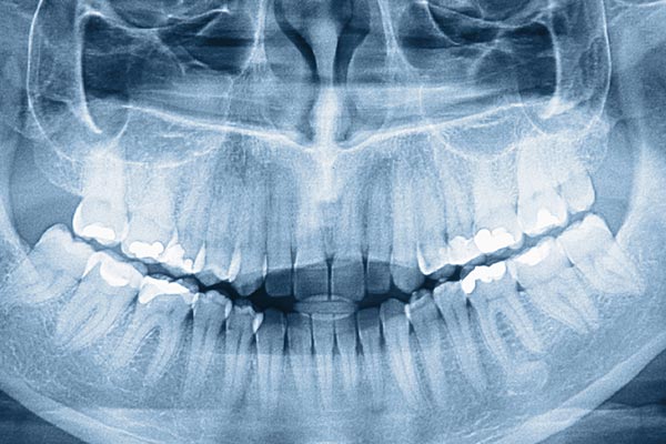
Modern orthodontic care begins with seeing the problem clearly. High-resolution digital imaging replaces traditional film with sensitive electronic detectors that capture fine detail in a fraction of the time. These images appear on-screen almost instantly, allowing your clinician to evaluate tooth positions, root alignment, and jaw relationships during the same appointment rather than waiting for film to develop. Faster imaging means quicker decisions and a more informed conversation about treatment options.
Because digital images are captured and stored electronically, they can be enhanced and reviewed from multiple angles without degrading the original data. Adjustments such as contrast enhancement and zooming help reveal subtle features that support accurate diagnosis and efficient treatment planning. Digital imaging also supports multiple views at once, so the dentist and patient can look at the same image together and discuss findings in real time.
At Longo Dietz Orthodontics we pair high-quality sensors with streamlined workflows to ensure every diagnostic image contributes directly to excellent care. The clarity and immediacy of digital radiographs reduce uncertainty and help our team design treatment plans that are both predictable and tailored to each patient’s needs.
Traditional impressions with putty can be uncomfortable and messy. Intraoral scanners change that by capturing a detailed three-dimensional map of the teeth and bite using a small, handheld wand. As the wand moves around the mouth it records millions of points that sophisticated software stitches together into an accurate 3D model. The result is a digital impression that faithfully represents tooth surfaces, gum contours, and occlusion without the gag reflex or tacky materials.
Digital scans are immediately viewable on-screen, allowing patients and providers to inspect the model together and spot any areas that may require attention. Because the data is precise and complete, labs and appliance manufacturers can produce custom devices—retainers, aligners, or brackets—with a high degree of accuracy. This precision reduces the need for adjustments and contributes to a smoother overall treatment experience.
Beyond comfort, intraoral scanning speeds up clinical appointments and improves predictability. Digital impressions can be transmitted securely to partners, archived for future reference, and integrated into the broader digital record—streamlining care while maintaining a patient-friendly approach.
Once diagnostic images and digital impressions are collected, powerful 3D planning tools turn data into actionable treatment strategies. Treatment planning software allows clinicians to simulate tooth movements, evaluate final positions, and visualize jaw relationships before treatment begins. These simulations help set realistic expectations and provide a visual roadmap that informs appliance selection and appointment sequencing.
Modern planning platforms support collaboration across specialties, enabling orthodontists to coordinate with general dentists, oral surgeons, and labs when cases require multidisciplinary care. The ability to export precise digital files for appliance fabrication ensures that what is planned in software can be realized clinically. Planning in three dimensions also helps anticipate potential challenges, such as space limitations or bite interference, so adjustments can be made proactively.
This level of digital planning contributes to more predictable outcomes and a more transparent patient experience. Patients benefit from seeing clear visualizations of proposed changes, while clinicians gain tools that improve the efficiency and accuracy of care.
Patient safety is a core principle of contemporary orthodontics, and technology plays a central role in protecting health while delivering excellent care. Digital radiography typically requires far less radiation than conventional film-based techniques, thanks to sensitive detectors and optimized exposure settings. Lower doses mean routine imaging can be performed with confidence while still providing the diagnostic detail clinicians need.
Beyond reduced radiation, other technological advances target comfort and convenience. Shorter image-capture times, ergonomic scanning devices, and software that limits the need for repeat exposures all contribute to a more comfortable visit. Staff also follow calibrated protocols and regularly maintain equipment to ensure consistent performance and safe operation across every appointment.
Clear communication about safety measures helps patients understand the purpose and expected benefits of each scan. When patients know why an image is being taken and how it supports their care, the experience feels more transparent and reassuring.
Electronic imaging and digital impressions are more than tools for diagnosis—they become part of a secure, integrated patient record. Digital storage makes it easy to retrieve prior images, compare progress over time, and document outcomes objectively. This continuity supports long-term care, especially for patients whose treatment spans months or years.
Digital records also facilitate seamless collaboration. When coordination with other dental specialists or laboratory partners is needed, secure file transfer ensures that high-quality data reaches the right team members promptly. Sharing precise digital models and imaging minimizes back-and-forth and helps external partners produce appliances that fit the planned treatment goals.
Robust data management practices protect patient privacy while enabling efficient clinical workflows. Backup systems, controlled access, and standard protocols for handling images and models ensure that digital records remain both accessible to the care team and safeguarded against unauthorized access.
In summary, technology in modern orthodontics streamlines diagnosis, improves comfort, and enhances the predictability of treatment. From high-resolution imaging and intraoral scanning to sophisticated planning software and secure digital records, these tools work together to support better outcomes and a more patient-centered experience. If you’d like to learn more about how our practice uses technology to improve care, please contact us for additional information.
We use high-resolution digital radiography and advanced sensor technology to capture clear images of tooth roots, jaw relationships and overall oral anatomy. These images appear on-screen almost instantly, allowing clinicians to evaluate findings during the same appointment and discuss options with patients in real time. Digital imaging often includes panoramic or cephalometric views when needed to assess growth, alignment and skeletal relationships.
Digital images can be enhanced and reviewed from multiple angles without degrading the original data, which supports more accurate diagnosis and treatment planning. The immediate availability of images reduces the need for repeat exposures and helps the care team design predictable, individualized treatment sequences. At Longo Dietz Orthodontics we pair high-quality sensors with streamlined workflows so every diagnostic image directly informs excellent patient care.
Intraoral scanners capture a precise three-dimensional map of the teeth and bite using a small handheld wand, eliminating the need for traditional putty impressions in many cases. The scanning process is faster and more comfortable, reducing gag reflex and the mess commonly associated with impression materials. Scans are viewable on-screen immediately, so patients and providers can inspect the model together and confirm that the data is complete.
Digital impressions deliver a high level of accuracy for appliance fabrication, which reduces the need for manual adjustments and remakes. Files can be transmitted securely to laboratories or manufacturers for rapid production of retainers, aligners and other devices. Scanned models are also archived electronically, enabling easy comparison of progress over time and straightforward retrieval for future reference.
Three-dimensional planning tools turn diagnostic images and digital impressions into visual simulations of proposed tooth movements and final positions. These simulations allow clinicians to evaluate outcomes, anticipate potential challenges such as space limitations or bite interference, and choose appliances and appointment sequencing that support predictable results. Planning software also helps set realistic expectations by showing patients clear visualizations of proposed changes.
Modern platforms support collaboration across specialties and enable the export of precise digital files for appliance fabrication, which helps ensure that what is planned in software can be realized clinically. The ability to coordinate with general dentists, oral surgeons and labs reduces delays and improves treatment efficiency. Overall, 3D planning contributes to more consistent outcomes and a more transparent patient experience.
Digital radiography typically requires far less radiation than conventional film-based techniques because of highly sensitive detectors and optimized exposure settings. Lower doses mean routine imaging can be performed with confidence while still providing the diagnostic detail clinicians need to plan care safely and effectively. Staff follow calibrated protocols and maintain equipment regularly to ensure consistent performance and minimal exposure.
Beyond lower doses, shorter capture times and fewer repeat images also contribute to safety and patient comfort. Clinicians order imaging based on clinical need and explain the purpose and benefits of each scan so patients understand how it supports their care. For children and other sensitive patients, exposure settings are adjusted appropriately to follow best-practice safety guidelines.
Digital imaging and intraoral scans become part of a secure, integrated patient record that is stored using encrypted systems and controlled access protocols. Regular backups, audit logs and role-based permissions help ensure that records remain available to the care team while protected from unauthorized access. Secure file-transfer methods are used when sharing data with specialists or laboratories to maintain patient privacy.
Robust data management practices support continuity of care by making it easy to retrieve prior images and compare progress over time. Standard retention and disposal policies govern how long records are kept and how they are archived for future reference. Clear communication about privacy measures helps reassure patients that their information is handled responsibly.
Yes, high-quality digital scans provide the precise geometry manufacturers and labs need to fabricate aligners, retainers and custom appliances with excellent fit. The scanned data is converted into standard digital files that technicians use to design and produce devices, helping reduce manual adjustments and fit issues. Because the digital workflow yields consistent, repeatable results, it shortens the feedback loop between the office and the lab.
Digital workflows also make it easier to request refinements or additional appliances when necessary, since the original scan can be reprocessed or updated rather than repeating physical impressions. This precision-driven approach improves the overall patient experience by minimizing surprises and improving the first-fit accuracy of appliances. Quality control checks remain a standard part of the process to confirm fit and function before delivery.
Advanced imaging, digital planning and precise appliance fabrication can improve treatment efficiency and reduce some sources of delay, but total treatment time depends on individual biology, the complexity of the case and patient compliance. Technology helps clinicians design more predictable movement sequences and can reduce the need for adjustments that stem from poor fit or inaccurate impressions. In many cases, better planning and manufacturing lead to smoother progress and fewer corrective visits.
Remote monitoring tools and digital communication can sometimes reduce routine in-office checkups by allowing the team to verify progress between appointments, but periodic clinical evaluations remain important for safe, effective care. Your clinician will explain how technology is used to streamline your specific treatment plan and will recommend an appointment schedule that balances efficiency with clinical oversight. Expectations about visit frequency and timeline are best set together during the planning phase.
Many technological advances directly reduce discomfort by shortening procedure times and eliminating unpleasant materials or techniques. Intraoral scanning removes the need for putty impressions, digital imaging shortens exposure and capture times, and modern bracket systems are lower profile and designed to reduce friction. Additional tools, such as the Dental Pain Eraser and other comfort-focused devices, are used in some offices to manage sensitivity without needles or gels when clinically appropriate.
Ergonomic instruments and streamlined workflows also help minimize chair time and the likelihood of repeat procedures, which contributes to a more comfortable overall experience. Clinicians combine technology with careful technique and clear communication to address patient concerns and make visits as pleasant as possible. Patients are encouraged to discuss comfort preferences so the team can tailor care accordingly.
Standardized digital files such as STL and DICOM enable secure, accurate sharing of images and models with general dentists, oral surgeons and laboratory partners. This interoperability allows specialists to review the same high-quality data, participate in joint treatment planning and fabricate appliances that match the planned outcome. Efficient file exchange reduces back-and-forth, speeds fabrication and supports consistent execution of multidisciplinary treatment plans.
Collaborative features in planning software let clinicians annotate images, simulate surgical or orthodontic sequences and discuss complex cases with clarity. When specialists are aligned on a digital plan, the clinical team can coordinate appointment sequencing and appliance delivery more effectively. These practices improve predictability and reduce delays that can arise from miscommunication or incompatible formats.
If you would like additional information about the digital tools and protocols we use, please contact our Omaha or Bellevue office to speak with a team member who can explain specific technologies and how they apply to your care. Our Omaha office can be reached at (402) 496-9733 and our Bellevue office can be reached at (402) 291-0528. Staff can describe the role of imaging, scanning and planning software in your treatment and arrange a demonstration when appropriate.
During new patient visits the clinical team reviews diagnostic results and walks through the digital records and simulation tools so patients understand their treatment path. We welcome questions and will provide clear explanations of safety measures, data handling practices and how technology is used to support comfort and predictability. This transparent approach helps patients make informed decisions about their orthodontic care.